Clinically grounded
Research questions begin with biological and clinical relevance.
Medical imaging · Neuroimaging · Artificial intelligence
I am Mingshi Chen (陈明诗), a PhD researcher at Amsterdam UMC. My work combines medical imaging, machine learning, and clinical knowledge to study brain health and build methods that remain useful beyond the model itself.
About
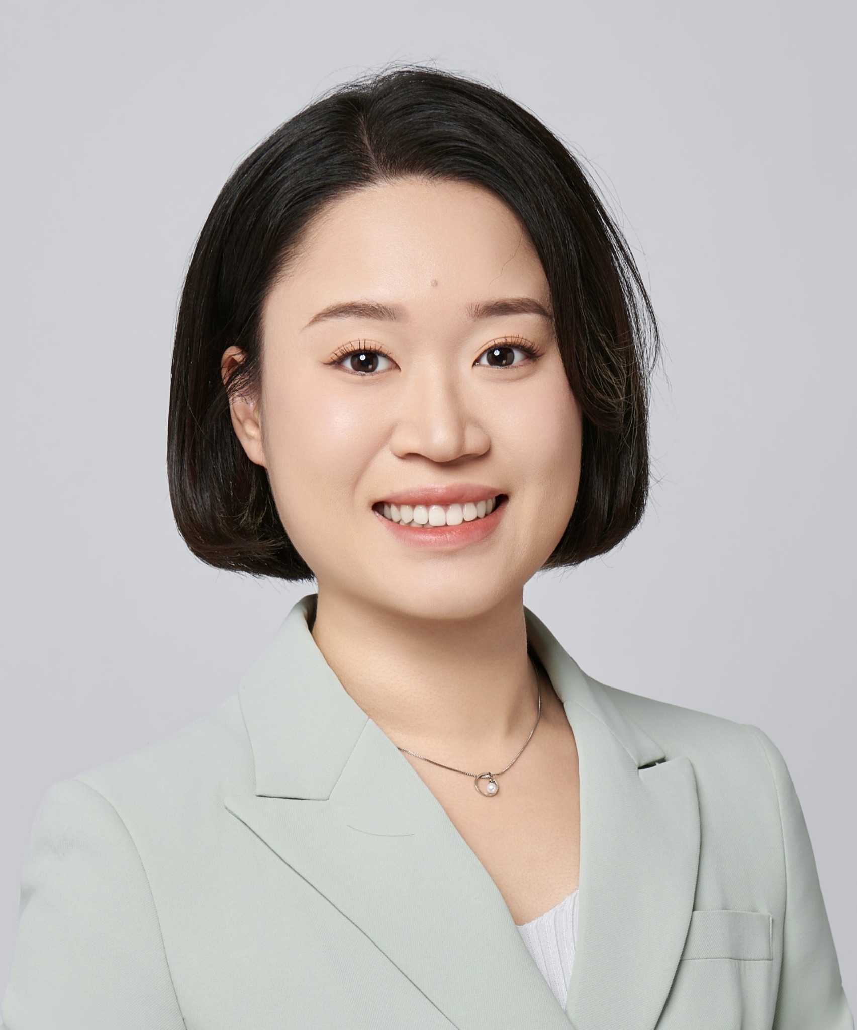
Mingshi Chen
陈明诗
PhD Researcher · Amsterdam UMC
My background spans clinical medicine, medical imaging, and data science. That combination shapes the questions I ask: not only whether a model performs well, but whether its output is interpretable, reproducible, and meaningful for patients and researchers.
At Amsterdam UMC, I work with structural MRI, diffusion MRI, functional MRI, radiomics, brain-age modelling, and longitudinal statistics. I enjoy building complete research pipelines—from imaging quality control and feature engineering to validation, visualization, and scientific communication.
Research questions begin with biological and clinical relevance.
Validation, sensitivity analysis, and clear limitations are part of the result.
Complex analyses should become understandable evidence, not a black box.
Research themes
My projects connect quantitative neuroimaging with cognition, psychiatric treatment, substance exposure, and individual differences.
Evaluating multiple grey- and white-matter brain-age models and asking how language experience relates to apparent brain ageing.
Studying structural brain changes before and after incident XTC exposure, including regional volumes, whole-brain patterns, dose, and memory.
Testing whether early clinical and task-fMRI signals can improve treatment outcome prediction in major depressive disorder.
Selected work
Peer-reviewed publication · Scientific Reports
A preregistered EMBARC analysis comparing clinical, regional mean-activation, radiomic, and whole-brain task-fMRI features for prediction of sertraline and placebo outcomes.
Peer-reviewed publication · NeuroImage: Clinical
A secondary analysis of a randomized clinical trial evaluating conventional MRI measures and radiomics with nested cross-validation in stimulant-naive children and adults with ADHD.
Book chapter · Academic Press
An overview of radiomics, machine learning, representation learning, and their application to extracting clinically useful information from magnetic resonance imaging.
Peer-reviewed publication · Cancer Imaging
Contributed to multimodal metabolic imaging research in primary central nervous system lymphoma.
Peer-reviewed publication · Cancers
Contributed to MRI-based characterization of primary central nervous system lymphoma.
Peer-reviewed publication · Radiotherapy & Oncology
Contributed to work comparing complementary imaging approaches in recurrent retropharyngeal disease.
Manuscript
Examines structural MRI-derived brain age across bilingual phenotypes with different levels of professional language experience and language-control demand.
PhD research
A prospective NeXT-cohort study combining behavioral replication, predefined hippocampal and thalamic analyses, baseline-derived PCA, mixed-effects modelling, and cumulative XTC dose.
Collaborative manuscript
A collaborative application of quantitative MRI and predictive modelling to sports-injury outcome.
For the complete and most current publication record, see Google Scholar or download my CV.
Experience & toolkit
2022 — present
Neuroimaging, medical AI, psychiatric imaging, longitudinal modelling, scientific writing, and student supervision.
2014 — 2022
BMed training in clinical medicine followed by an MClinMed in imaging and nuclear medicine at Sun Yat-sen University and SYSU Cancer Center.
Imaging
Structural MRIDTIfMRI FreeSurferFastSurferFSLData science
PythonPyTorchMONAI scikit-learnstatsmodelsRAnalysis
RadiomicsBrain ageMixed models Cross-validationVisualizationContact
I am interested in postdoctoral research and medical-AI roles involving neuroimaging, model validation, clinical translation, or multimodal data.